

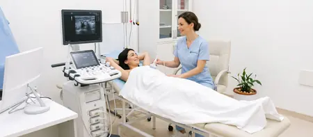
What a breast ultrasound examines
A breast ultrasound shows the breast layer by layer: the skin, fatty tissue, glandular tissue and milk ducts. The doctor scans the whole surface of both breasts, including the area around the nipple.
The scan also covers the armpit, where the lymph nodes that drain the breast are located. An enlarged or changed node in the armpit is sometimes the first sign worth checking.
The main strength of ultrasound is that it clearly tells a fluid-filled cyst from a solid lump. That is the key question with almost every lump you can feel. Since there is no radiation, the scan can be repeated as often as needed, which makes it well suited for regular follow-up.
When a breast ultrasound is done
A breast ultrasound is done when there is a specific complaint and also as a routine check-up for women without symptoms. A breast lump is a reason to get checked without delay, even if it does not hurt. Most lumps turn out to be benign, but only an examination can confirm that.
The most common reason is a lump or thickening you can feel in the breast or armpit. The scan is also done for breast pain or tenderness that does not follow your menstrual cycle. Nipple discharge is another reason, especially if it is bloody or from one breast only. The same applies to an inverted nipple or a change in the skin of the breast.
Ultrasound complements a mammogram, especially with dense breast tissue, and is used to monitor a previously found cyst or fibroadenoma. Routine check-ups are advised for women under 40 and for women whose close relative had breast cancer. While breastfeeding, swelling, redness and pain in the breast are a reason for the scan.
Preparation and when in your cycle to have a breast ultrasound
No special preparation is needed for a breast ultrasound. You eat, drink and take your medication as usual.
Timing matters more. If you have periods, the scan is best done between day 5 and day 12 of your cycle, counting from the first day of bleeding. The glandular tissue is calmest then, the breasts are not tense and the image is clearest. Before a period the tissue swells, which can hide small changes or create the impression of new ones.
Women after menopause, pregnant women and breastfeeding mothers can come on any day. If you have found a lump, do not wait for the ideal day of your cycle. Book the scan as soon as possible.
Before the scan it helps to:
- bring earlier breast ultrasound and mammogram reports, with images if you have them
- note the first day of your last period
- wear a two-piece outfit, since the scan is done without a top
- tell the doctor if anyone in your family has had breast or ovarian cancer
Breast ultrasound or mammogram: the difference
Ultrasound and mammography do not replace each other. Each method sees what the other can miss, so they are often done together.
A mammogram is an X-ray of the breasts. It is good at detecting microcalcifications, tiny calcium deposits that can be an early sign of disease. That is why it is the standard screening method for women over 40 or 50, as advised by their doctor.
Ultrasound uses no radiation and shows dense, glandular breast tissue better. Younger women usually have dense breasts, which look white on a mammogram, so changes are harder to spot. For this reason ultrasound is usually the first examination for women under 40.
For older women ultrasound complements the mammogram. The doctor recommends it when a mammogram shows an unclear shadow, when the breast is dense or when it needs to be checked whether a finding is a cyst or a solid mass.
What a breast ultrasound is like
You lie on your back with your arm raised above your head. In this position the breast tissue spreads evenly over the chest and is easier to examine.
The doctor applies gel and slowly moves the probe over the whole breast, then over the armpit. If you have felt a lump, show the doctor the exact spot before the scan begins. The doctor measures any suspicious change in three directions and checks how it behaves under the pressure of the probe.
The scan does not hurt. If your breasts are tender, tell the doctor and the pressure will be lighter. Both breasts and armpits are examined in 15 to 20 minutes, and there are no restrictions afterwards.
What the report says: BI-RADS categories
The doctor describes each finding: size, shape, margins and position, usually by clock-face position and distance from the nipple. This makes it easy to find the same spot at the next check-up.
The most common findings are cysts and fibroadenomas, and both are benign. A cyst is a small fluid-filled sac, common between the ages of 35 and 50. A fibroadenoma is a firm, mobile nodule seen most often in younger women.
The doctor tells you the main conclusions at the end of the scan, and the written report follows shortly. Images are stored digitally, so every check-up can be compared with the previous one.
The report ends with a BI-RADS category. This is an international scale from 0 to 6 that briefly states how suspicious a finding is and what the next step should be:
- BI-RADS 0: the scan is not enough for a conclusion and further imaging is needed, for example a mammogram
- BI-RADS 1 and 2: normal findings, or a finding that is certainly benign, for example a simple cyst
- BI-RADS 3: a probably benign finding, with a follow-up scan advised in about six months
- BI-RADS 4 and 5: a suspicious finding, and the doctor recommends a biopsy
- BI-RADS 6: the diagnosis has already been confirmed by biopsy and the scan is used for monitoring
Breast ultrasound during pregnancy and breastfeeding
A breast ultrasound is safe during pregnancy and breastfeeding because it uses no radiation. In this period it is usually the first and often the only examination needed.
Hormones during pregnancy and breastfeeding change the breast: the tissue grows and becomes denser, and the milk ducts widen. This makes a new lump harder to feel and harder to judge by self-examination. A lump that does not go away within one to two weeks should be checked.
During breastfeeding ultrasound helps with breast inflammation (mastitis). It shows whether an abscess has formed, a collection of pus that antibiotics alone do not resolve. It also shows a galactocele, a cyst filled with milk. High fever, redness and severe breast pain need a doctor the same day.
Feed your baby or express milk before the scan, because an emptied breast is easier to examine.
Breast ultrasound cost
An ultrasound of both breasts with an armpit check costs 6,000 RSD. The price includes the scan, the specialist's interpretation and a written report.
If the doctor recommends a further examination, for example a mammogram or a surgical consultation, it is arranged separately.
- Ultrasound of both breasts
- Check of the armpit lymph nodes
- Written report with BI-RADS category
Breast ultrasoundfrom 6,000 RSD
View the current price in our price listFor an appointment call 062-399-888.
Frequently asked questions about breast ultrasound
Ideally between day 5 and day 12 of your cycle, counting from the first day of your period. The breasts are not tense then and the image is clearest. If you have found a lump, do not wait for this window and book the scan straight away.
Neither is better in every respect, because they show different things. For women under 40 ultrasound is usually the first choice because of dense breast tissue. For older women the mammogram is the standard screening test and ultrasound complements it.
No. You only feel cool gel and light pressure from the probe. If your breasts are tender before your period, tell the doctor and the pressure will be gentler.
A scan of both breasts and armpits takes 15 to 20 minutes. If a finding needs detailed measurement, it may take a little longer.
At Medical Time hospital a breast ultrasound costs 6,000 RSD. The price covers both breasts and armpits and a written report from the specialist.
BI-RADS 3 means the finding is probably benign. The doctor usually recommends a follow-up ultrasound in about six months to see whether it changes.
Yes, and it is safe because there is no radiation. Breastfeeding mothers should feed the baby or express milk before the scan, as the breast is then easier to examine.
Yes. Men have them for breast enlargement (gynecomastia), a lump or pain under the nipple. The scan is the same as for women.
Related pages
Examinations often done alongside a breast ultrasound