

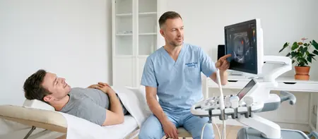
What a scrotal ultrasound examines
The scrotum is the skin sac that holds the testicles, the epididymis and the spermatic cords. That is why the same examination is called both a testicular ultrasound and a scrotal ultrasound. The doctor scans both sides and compares them, because the difference between left and right is often the most important finding.
The image shows the size and structure of each testicle, the epididymis next to it, any fluid around the testicle and the veins of the spermatic cord. Ultrasound reliably shows whether a lump is inside the testicle or next to it. Every further step depends on that distinction.
Color Doppler is the part of the ultrasound scan that shows blood flow in color. In the scrotum it shows whether the testicle gets enough blood, whether the tissue is inflamed and whether the veins are enlarged. That is why patients often ask for a testicular ultrasound with Doppler.
When a testicular ultrasound is done
A testicular ultrasound is the first examination for almost any change in the scrotum. Any firm lump on a testicle needs to be checked without delay, even if it does not hurt. Testicular cancer is the most common cancer in men between 15 and 35, and when found early it has good chances of successful treatment. A monthly self-check is useful, and any new change is a reason for an ultrasound.
The most common reasons for the scan are:
- a lump, thickening or hard spot on a testicle
- swelling or enlargement of one side of the scrotum, or pain and swelling after an injury or infection
- a dull ache or heavy feeling in the scrotum, especially after standing for a long time
- enlarged veins above the testicle that feel like a bag of worms
- difficulty conceiving and an abnormal semen analysis, or a check-up after varicocele or hydrocele surgery
Sudden severe testicular pain is an emergency
Sudden, severe pain in one testicle, especially in boys and young men, can be testicular torsion. The spermatic cord twists and cuts off the blood supply to the testicle. The testicle can only be saved if it is operated on within the first few hours.
If the pain starts suddenly, if the testicle is swollen or sits higher than usual, or the pain comes with nausea and vomiting, do not book a routine scan. Call an ambulance on 194 straight away or go to the nearest emergency department.
Pain that builds up gradually over several days, with swelling and fever, is more often an inflammation of the epididymis (epididymitis). It also needs to be seen the same or the next day. Ultrasound with Doppler then shows increased blood flow in the inflamed tissue.
Preparing for a testicular ultrasound
No preparation is needed for a testicular ultrasound. You eat, drink and take your medication as usual.
Shower before the scan and wear comfortable clothing. Bring earlier ultrasound reports, semen analyses and hormone test results if you have them. If you have had surgery in the groin or scrotum, bring the discharge summary as well.
If you also need an abdominal scan, ask about the combined abdominal and scrotal ultrasound. For that examination you come fasting, because the abdominal part requires it.
What a scrotal ultrasound is like
You lie on your back. The doctor places a folded towel under the scrotum and covers the penis, so that the testicles stay still and you feel more comfortable. The doctor then applies gel and gently moves the probe over both sides.
To check for a varicocele, the doctor will ask you to take a deep breath and bear down as if straining (the Valsalva maneuver). Part of the scan is sometimes done standing up, because enlarged veins fill with blood better in that position.
The scan does not hurt. The probe pressure is light, and the doctor takes extra care if the testicle is painful from inflammation. A scrotal ultrasound usually takes about 15 minutes, and you can go back to your day straight after.
What a testicular ultrasound report shows
The doctor describes the size and structure of both testicles, the appearance of the epididymis, the amount of fluid in the scrotum and the width of the veins. You hear the main conclusions at the end of the scan, and the written report follows shortly.
Images are stored digitally, so at a follow-up it is easy to see whether a varicocele or cyst has changed.
The most common findings are:
- varicocele: enlarged veins of the spermatic cord, most often on the left. The report states the vein width and whether blood flows back when you strain.
- hydrocele (fluid around the testicle that enlarges the scrotum) and an epididymal cyst or spermatocele beside it, usually harmless
- epididymitis and orchitis: inflammation of the epididymis or testicle, with increased flow on Doppler
- microcalcifications: tiny bright spots in the testicle that are usually just monitored
- a solid lesion inside the testicle: needs a prompt urology review and blood tests
After the scan: treating varicocele and hydrocele
A small varicocele without symptoms and with a normal semen analysis is usually just monitored. Surgery is suggested when the veins cause pain, when the testicle shrinks or when there are problems conceiving. The procedure is described on the varicocele surgery page.
In adults a hydrocele rarely goes away on its own. If it is large, gets in the way when walking or keeps growing, it is treated surgically. You can read more on the hydrocele surgery page.
The decision on treatment is made by a urologist, based on the ultrasound, a physical examination and blood tests if needed. At Medical Time the urologist, the laboratory and surgery are in the same hospital, so you can book the next step straight away.
Testicular ultrasound cost
A testicular and scrotal ultrasound costs 6,000 RSD. The price includes the scan of both sides, the specialist's interpretation and a written report.
If you also need an abdominal scan, ask about the combined abdominal and scrotal ultrasound. We will confirm at booking whether your scan includes a Doppler flow assessment.
- Ultrasound of both testicles and the scrotum
- Assessment of varicocele and hydrocele
- Written specialist report
Testicular ultrasoundfrom 6,000 RSD
View the current price in our price listFor an appointment call 062-399-888.
Frequently asked questions about testicular ultrasound
Yes, these are two names for the same examination. The scrotum is the sac that holds the testicles, so the scan covers the testicles, the epididymis and the surrounding veins.
No special preparation is needed. Just shower beforehand and bring earlier reports if you have them.
No. You feel the gel and light pressure from the probe. If the testicle is painful from inflammation, the doctor uses even less pressure.
Usually about 15 minutes. If a varicocele is also checked standing up, the scan takes a few minutes longer.
At Medical Time hospital a testicular and scrotal ultrasound costs 6,000 RSD. The price covers both sides and a written report from the specialist.
It is an ultrasound combined with color Doppler, which shows blood flow in color. It reveals enlarged veins in varicocele, increased flow in inflammation and weak or absent flow in testicular torsion.
Yes, ultrasound with Doppler is the main test for varicocele. It shows the width of the veins and whether blood flows back when you bear down.
Call an ambulance on 194 straight away or go to an emergency department. Sudden, severe pain can be testicular torsion, which needs surgery within the first few hours.
Related pages
Related examinations and treatment