

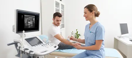
What a soft tissue ultrasound is and what it covers
Soft tissues are everything between the skin and the bone: the fat layer under the skin, muscles, tendons, lymph nodes and the abdominal wall. For this scan the doctor uses a high-frequency linear probe. It does not reach deep, but it shows structures near the surface in fine detail, down to a few millimetres.
The scan answers the question that usually brings you in: what is this lump. The doctor sees whether it is filled with fluid or solid, whether its edges are clear, how large it is and whether blood vessels run through it. This is how harmless changes are told apart from those that need further tests.
The scan is targeted at the spot that worries you. If the lump is on the neck, the doctor also checks the lymph nodes on both sides of the neck. Knee and shoulder scans have their own pages, because there the focus is on tendons, ligaments and fluid in the joint.
When it is done: lumps, lymph nodes, muscles and hernias
The most common reason is a lump you found yourself or one your doctor noticed. Ultrasound is usually the first test for such a change, before any procedure. It is also used to follow up a change that has been monitored before.
- a soft or firm lump under the skin anywhere on the body, or suspected lipoma or sebaceous cyst before removal
- enlarged lymph nodes in the neck, armpit or groin, or a neck lump that does not belong to the thyroid
- muscle pain or swelling after a blow or strain, with a suspected tear or haematoma
- a bulge in the groin, around the navel or at a surgical scar, with a suspected hernia
- redness, warmth and swelling that may be an abscess
Lymph node ultrasound: neck, armpit, groin
Lymph nodes swell after a cold, a sore throat, a tooth problem or a skin wound, and they usually shrink within two to four weeks. Ultrasound shows the size and shape of the node and whether its normal inner structure is preserved. An oval node with a visible central part (the hilum) is most often reactive, meaning harmless.
See a doctor without delay if the node is hard, does not move, keeps growing or has not gone away after three weeks. The same applies if the swollen node comes with fever, night sweats or weight loss you cannot explain.
Neck ultrasound
During a neck ultrasound the doctor examines the lymph nodes, the salivary glands below the jaw and in front of the ear, the muscles and the tissue under the skin. When the lump is in the thyroid area, a thyroid ultrasound is done instead, and a carotid Doppler is used for the neck blood vessels.
What ultrasound shows: lipoma, sebaceous cyst, cyst, hernia
Each common change has a typical look. The doctor recognises it during the scan and explains what they see right away.
A lipoma is a soft, mobile lump of fatty tissue with clear edges and no increased blood flow. Cysts have contents: a sebaceous (epidermoid) cyst holds thick material, while a ganglion and other cysts hold fluid. A sebaceous cyst is round and attached to the skin, sometimes with a visible pore. A ganglion most often sits next to the tendons of the hand and foot.
For a lymph node, the doctor measures its size, assesses its shape and says whether it is reactive or altered. An abscess is a collection of pus with inflamed surrounding tissue. After an injury the scan shows a haematoma, which is pooled blood, or broken muscle fibres in a tear. An abdominal wall hernia is an opening through which fat or bowel pushes out.
Ultrasound of an abdominal wall hernia
When a hernia is suspected, the doctor scans you lying down and, if needed, standing. You will be asked to cough or tense your stomach, because that is when the hernia appears and the size of the opening can be measured. The groin, the navel, the midline above the navel and old surgical scars are all checked.
If a hernia suddenly becomes hard and very painful, cannot be pushed back in and you feel sick or vomit, call emergency services. This may be a strangulated hernia, and it should not wait for a scheduled scan.
Preparation and what happens during the scan
No preparation is needed for a soft tissue ultrasound. Eat, drink and take your medication as usual. Wear clothing that easily uncovers the area, and if you have had an earlier scan of the same lump, bring the report so the doctor can compare the size.
The doctor applies gel and gently moves the probe over the lump and the area around it. They compare it with the same spot on the other side of the body and may switch on Doppler to see blood flow through the lump. The scan is painless. Over an inflamed lump the pressure of the probe can feel uncomfortable, so feel free to ask the doctor to press more lightly.
A scan of one area takes 15 to 20 minutes. If you have several lumps in different places, mention it when booking so we can allow enough time.
What happens after the results
For most lumps the result is reassuring: a lipoma, a sebaceous cyst or a reactive lymph node. A lipoma or sebaceous cyst is removed only if it bothers you, grows or becomes inflamed, and removal is a simple surgical procedure. A reactive node is usually just checked again after a few weeks.
If a change does not look typical, the doctor may recommend blood tests, a follow-up scan, a surgical consultation, or a needle aspiration and biopsy. At Medical Time the laboratory, surgeons and other specialists work in the same hospital, so the next step can be booked right away. You receive the written report shortly, and the images are stored digitally for comparison at follow-up.
Soft tissue ultrasound cost
A soft tissue and muscle ultrasound costs 6,000 RSD and covers one area with a specialist's interpretation. The current price from our price list is shown below.
- Soft tissue and muscle ultrasound
- Ultrasound of lymph nodes in the neck, armpit or groin
- Ultrasound of a lump, lipoma or sebaceous cyst
Soft tissue ultrasoundfrom 6,000 RSD
View the current price in our price listFor an appointment call 062-399-888.
Questions about soft tissue ultrasound
A soft tissue ultrasound at Medical Time costs 6,000 RSD. The price covers one area and the specialist's interpretation.
No, no preparation is needed. You can eat and drink normally; just wear clothing that easily uncovers the area being scanned.
Yes, in most cases it can. A lipoma is a mobile lump of fatty tissue, while a sebaceous cyst is attached to the skin and filled with thick content, so they look different on screen.
A scan is advisable when the node has not gone away after three weeks, keeps growing, is hard or does not move. It is also needed if the node comes with fever, night sweats or weight loss.
Yes, ultrasound shows an abdominal wall hernia and the size of the opening. The doctor asks you to cough or tense your stomach, because the hernia is easiest to see then.
No, the scan is painless. Only over an inflamed or freshly injured area can the probe feel uncomfortable, and the doctor then presses more lightly.
Yes. Ultrasound uses no radiation, so it is safe for children and in pregnancy; in children it is most often used for neck lymph nodes.
For most lumps near the surface ultrasound is enough. The doctor recommends an MRI when the change is deep, large or still unclear after the ultrasound.
Related pages
Related exams and specialists: