

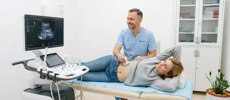
What a kidney and urinary tract ultrasound covers
During a kidney ultrasound the doctor examines both kidneys, the ureters and the bladder. They measure the size of the kidneys and the thickness of the kidney tissue (parenchyma), which becomes thinner in chronic kidney disease.
In the centre of each kidney is the collecting system, where urine gathers. When urine cannot drain, for example because of a stone, this system widens. That widening (hydronephrosis) is clearly visible on ultrasound.
Healthy ureters are thin and hard to see. They become visible when they are widened, which is useful information in itself. The doctor looks at the bladder when it is full: its wall, its contents and how much urine is left after you pass water. In men the doctor can also estimate the size of the prostate through the abdomen.
When a kidney ultrasound is done
A kidney ultrasound is the first test for most urinary symptoms and for pain in the flank.
Severe flank pain with high fever and chills, or a complete inability to pass urine, needs emergency care. In that case call 194, the emergency number in Serbia.
The most common reasons are:
- pain in the flank or back, especially cramping pain that spreads to the groin
- blood in the urine, visible or found in a urine test
- frequent urinary tract infections, difficulty passing urine, a weak stream or a feeling that the bladder is not empty
- raised creatinine or urea in blood tests, or high blood pressure, particularly in younger people
- monitoring the kidneys in diabetes or long-standing hypertension, or follow-up of a stone, a cyst or treatment
How to prepare for a kidney ultrasound
You do not need to fast for the kidneys alone. The bladder, however, is only clearly visible when it is full, and a kidney and urinary tract ultrasound usually includes it.
Take your regular medication as usual unless your doctor says otherwise.
Before the scan:
- an hour before the scan, drink half a litre to a litre of still water
- then do not pass urine until the doctor says so, and tell reception if holding it is hard
- the day before, avoid fizzy drinks and foods that cause bloating
- if the abdomen or a renal artery Doppler is scanned as well, fast for 6 to 8 hours
- bring your urine test, blood tests and earlier scans
How a kidney ultrasound is done
The scan starts with the bladder while it is full. You lie on your back, and the doctor applies gel to the lower abdomen and examines the bladder.
You then turn onto your side or front, because the kidneys sit at the back, under the ribs. The doctor asks you to take a deep breath so the kidney moves down below the ribs and is easier to see.
At the end the doctor may ask you to empty your bladder and then look at it again. This measures how much urine is left after passing water (post-void residual).
A kidney ultrasound takes 15 to 20 minutes and does not hurt. If a kidney is inflamed or a stone is moving, pressure from the probe on the flank can be uncomfortable.
Kidney stones and other findings
A kidney stone appears on ultrasound as a bright spot with a shadow behind it. The doctor notes where it is and how big it is, because size determines whether the stone can pass on its own.
Ultrasound sometimes misses a small stone in the ureter, but it shows the widening of the collecting system above it. When treatment planning requires it, a urologist may also request a CT scan.
Besides stones or gravel, the report often describes simple cysts, which are common after 50 and usually harmless. It also notes a dilated collecting system (hydronephrosis), or a small kidney or thinned parenchyma in chronic disease. Sometimes the scan shows a lesion in the kidney that needs further tests.
In the bladder the doctor describes a thickened wall, a stone or retained urine, and in men an enlarged prostate. The doctor tells you the main conclusions during the scan, and you receive the written report shortly afterwards. The report and images are stored digitally, so the size of a stone or cyst can be compared from one check-up to the next.
Kidney ultrasound in children
Ultrasound is the first kidney test in children, because it uses no radiation and does not hurt. A small child can sit on a parent's lap or lie next to them.
In children it is most often done for repeated urinary tract infections or a fever without a clear cause. It is also done when a pregnancy ultrasound showed a dilated collecting system. Other reasons are bedwetting after the age of five, abdominal pain or blood in the urine.
Babies cannot hold a full bladder, so the doctor examines the bladder whenever it happens to be full. Older children drink water according to their age. When you book, tell us how old your child is so you get exact instructions.
Renal artery Doppler
A renal artery Doppler shows blood flow through the arteries that supply the kidneys. Narrowing (stenosis) of these arteries reduces blood flow to the kidney and can cause high blood pressure that is hard to control with medication.
It is done when blood pressure stays high despite several medicines, when hypertension starts at a young age, when the kidneys differ in size or when creatinine rises after starting certain blood pressure medicines.
For a renal artery Doppler you fast for 6 to 8 hours, because bowel gas hides the blood vessels. The scan takes longer than a kidney ultrasound alone. We do it in combination with an abdominal ultrasound, for 6,000 RSD.
Kidney ultrasound cost
A kidney and urinary tract ultrasound costs 6,000 RSD, including the specialist's report. A renal artery Doppler is done together with an abdominal ultrasound, also for 6,000 RSD.
- Ultrasound of the kidneys and ureters
- Abdominal ultrasound with renal artery Doppler
Kidney ultrasoundfrom 6,000 RSD
View the current price in our price listFor an appointment call 062-399-888.
Kidney ultrasound: frequently asked questions
A kidney and urinary tract ultrasound at Medical Time costs 6,000 RSD. A renal artery Doppler is done together with an abdominal ultrasound, also for 6,000 RSD.
Not for the kidneys alone. You need a full bladder: an hour before the scan drink half a litre to a litre of water and do not pass urine. You fast only if the abdomen or a Doppler is scanned as well.
You lie on your back and then on your side, while the doctor moves a probe with gel over your abdomen and flanks. The full bladder is examined first, then the kidneys. The scan takes 15 to 20 minutes.
Yes, kidney stones show up well on ultrasound, and the doctor also measures their size. A very small stone in the ureter can be missed, but the widening it causes is visible.
No. You feel the gel and the pressure of the probe. A full bladder can be uncomfortable, but you empty it right after that part of the scan.
Yes, ultrasound is safe even for newborns because there is no radiation. The amount of water before the scan is adjusted to the child's age.
Ultrasound shows the structure of the kidneys: size, stones, cysts and dilation. Doppler measures blood flow through the kidney arteries and looks for narrowing, usually because of high blood pressure.
It depends on the result. Stones, the prostate and the bladder are the field of a urologist, while reduced kidney function and blood pressure belong to an internist. The doctor who performs the ultrasound will recommend the next step.
Related pages
Related exams and doctors