

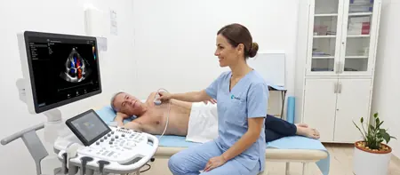
What a heart ultrasound is and what it shows
Heart ultrasound, echocardiography and "echo" are names for the same test. The doctor places a probe on your chest, and the machine turns reflected sound waves into a moving image of your beating heart.
An ECG records the heart's electrical activity. An ultrasound shows its structure and how the muscle moves. The doctor sees the size of the atria and ventricles, the thickness of the walls and how they contract with every beat.
Colour Doppler of the heart is part of the same scan. It shows in colour how blood moves through the chambers and valves. This reveals whether a valve leaks and lets blood flow backwards, or is too narrow so blood passes with difficulty.
The scan shows chamber size, wall thickness and pumping strength, expressed as the ejection fraction. The doctor assesses all four valves: mitral, aortic, tricuspid and pulmonary. The first part of the aorta and any fluid in the sac around the heart (pericardium) are also visible. The image also allows an estimate of pressure in the lung circulation.
When an echocardiogram is done
An echocardiogram is usually recommended by a cardiologist or a GP when symptoms or test results point to the heart. It answers two questions: is the heart structurally healthy, and how well does it pump. It is also used to assess the heart before surgery or before starting sport.
The most common reasons for a heart ultrasound are:
- a heart murmur the doctor heard with a stethoscope
- shortness of breath on exertion or when lying flat, swollen legs and getting tired quickly
- high blood pressure over many years
- palpitations, skipped beats or changes on an ECG
- a check-up after a heart attack or heart surgery, and monitoring known valve disease or heart failure
Heart murmur
A murmur is the sound of blood flowing faster or swirling inside the heart. In many people it is harmless. A heart ultrasound shows whether the murmur comes from a valve problem or a hole in the heart wall, or whether the heart is healthy.
After a heart attack
After a heart attack, part of the left ventricle wall may move less well. An echocardiogram shows how large that area is and what the ejection fraction is. The cardiologist uses this to adjust treatment and plan follow-up visits.
Preparing for a heart ultrasound
No special preparation is needed. You can eat, drink and take your usual medication as normal.
Wear clothes you can easily take off to the waist. Bring previous heart ultrasound reports, ECGs, hospital discharge letters and a list of your medicines. With earlier results the doctor can see whether anything has changed.
How the scan works and how long it takes
You lie on your left side with your left arm raised behind your head. In this position the heart sits closer to the chest wall and the image is clearer. The doctor applies gel and scans the heart from several points: beside the breastbone, below the left breast, under the ribs and above the collarbone.
During the scan you will hear the sound of blood flow. This is a normal part of colour Doppler. The doctor may ask you to breathe out and hold your breath briefly, because air in the lungs hides the heart.
A heart ultrasound takes 20 to 30 minutes. It does not hurt. You only feel the cool gel and light pressure from the probe. Afterwards you can drive and go straight back to your day.
Your report: ejection fraction and valves
The echocardiogram report lists the measured dimensions of the heart, how the left and right ventricles work and a description of each valve. The specialist tells you the main findings during the scan, and you receive the written report shortly after. Images are stored digitally, so they can be compared at your next check-up.
Ejection fraction
The ejection fraction (EF) shows what percentage of blood the left ventricle pumps out with each beat. In adults, a value of about 50 to 70 percent is usually considered normal. A lower value points to weaker pumping and needs a cardiologist's assessment.
Valves
For each valve the report states whether it is normal, narrowed (stenosis) or not closing properly (regurgitation). Mild backflow through some valves is common in healthy people. The doctor grades any change as mild, moderate or severe.
When not to wait for an appointment
A heart ultrasound is a planned test. Severe chest pain or tightness, sudden breathlessness, fainting or a cold sweat need emergency care, not an appointment. Call the ambulance on 194 straight away.
Heart ultrasound cost
A heart ultrasound with colour Doppler at Medical Time costs 6,000 RSD. The price covers the echocardiogram, colour Doppler of blood flow through the heart and a written report. If you also want a cardiologist consultation with an ECG, mention it when booking and we will arrange the appointments so you come in as few times as possible.
- Heart ultrasound with colour Doppler (echocardiogram)
Heart ultrasoundfrom 6,000 RSD
View the current price in our price listFor an appointment call 062-399-888.
Questions about heart ultrasound
Yes, they are two names for the same test. Echocardiogram is the medical term, and people also say cardiac ultrasound or simply echo.
A heart ultrasound with colour Doppler at Medical Time costs 6,000 RSD. The written report is included.
No preparation is needed. Eat, drink and take your medication as usual, and bring any previous heart test results.
It takes 20 to 30 minutes and does not hurt. You only feel cool gel and light pressure from the probe on your chest.
An ECG records the heart's electrical activity and rhythm, while an ultrasound shows its structure, valves and pumping strength. The tests complement each other, so a cardiologist often asks for both.
A low ejection fraction means the left ventricle pumps less strongly than it should. A cardiologist determines the cause and treatment, together with your other results and symptoms.
Yes, an echocardiogram is safe in pregnancy because it uses no radiation. It is done when a doctor suspects a heart problem or because of an earlier heart condition.
Not directly. The coronary arteries are hard to see on ultrasound, but the scan shows whether part of the heart wall moves less well, which can point to reduced blood supply.
Related pages
Related tests and doctors