

What an ultrasound scan is and why it is safe
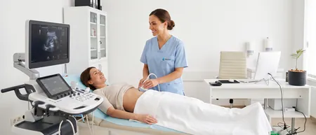
An ultrasound scan (sonography, echo) uses high-frequency sound waves that the human ear cannot hear. The probe sends them through the skin, they bounce off the organs, and the machine turns those echoes into a live image on the screen.
Unlike X-ray and CT, ultrasound uses no ionising radiation. The waves do not stay in the body and do not damage tissue, so the scan is safe for children, older people and pregnant women.
The image forms while the doctor moves the probe, so the specialist reads what they see during the scan itself. Ultrasound shows soft organs and fluid well, and is less useful for bone and air-filled organs such as the lungs and bowel.
Types of ultrasound we perform
An ultrasound is booked by body area. Below is a short description of each group, and every type of scan has its own page with preparation, the course of the scan and the price.
Abdomen and urinary tract
An abdominal ultrasound shows the liver, gallbladder, pancreas and spleen, and is most often done for pain, bloating or a liver check. A kidney and urinary tract ultrasound looks for stones, cysts and blocked urine flow.
Neck and thyroid
A thyroid ultrasound shows the size and structure of the gland and any nodules. It is done for an enlarged gland, abnormal hormone levels or a lump you can feel in the neck.
Breasts
A breast ultrasound tells a cyst from a solid lump and complements mammography. It is especially useful for younger women, whose breast tissue is denser.
Men: testicles and prostate
A testicular (scrotal) ultrasound is done for swelling, pain or a lump in the scrotum. A prostate ultrasound is done through the abdomen and shows the size of the prostate and how much urine stays in the bladder after you pass water.
Pelvis
A pelvic ultrasound is done through the abdomen and shows the bladder and, in women, the uterus and ovaries. The most common reasons are lower abdominal pain and irregular periods.
Joints and soft tissues
A soft tissue ultrasound shows lumps under the skin, lipomas, cysts and enlarged lymph nodes in the neck, armpit or groin. A knee or shoulder ultrasound shows tendons, ligaments and fluid in the joint after an injury or with long-standing pain.
Heart
A heart ultrasound (echocardiography) shows how well the heart pumps blood and how the heart valves work. It is done for breathlessness, a heart murmur, high blood pressure or after a heart attack.
Blood vessels: Doppler
A leg Doppler shows blood flow through the veins and arteries, varicose veins and blood clots. A carotid Doppler shows plaque and narrowing in the neck arteries that carry blood to the brain.
Home ultrasound
For bedridden patients and those who cannot easily travel, a doctor comes to your address in Belgrade with a portable machine. You agree on the area to be scanned when you book.
General preparation for an ultrasound
A thyroid, breast, soft tissue, joint or heart ultrasound needs no special preparation. An abdominal scan requires you to come fasting, and a bladder, prostate or pelvic scan requires a full bladder. Bring your previous reports and wear clothes that are easy to remove from the area being scanned. The exact instructions are on the page for your type of ultrasound, and you also get them when you book.
What the ultrasound scan is like
You lie on your back or side, and the doctor applies a clear gel to the skin and moves the probe over it. The gel feels cool and the probe presses lightly, with no pain and no needles. From time to time the doctor will ask you to take a deep breath or change position.
A scan of one area takes 10 to 30 minutes. Afterwards you wipe off the gel and go straight back to your day.
Your ultrasound report
The doctor tells you the main findings during the scan, and you receive the written report shortly afterwards. The report describes the size and appearance of the organs, any changes found and a recommendation for next steps.
Sometimes an ultrasound does not give a final answer. In that case the doctor suggests the next step, for example blood tests, a Doppler or a scan with another method.
Reports and images are stored digitally, so every follow-up can be compared with the previous scan. Laboratory tests, further diagnostics and specialist appointments can all be booked in one place, at the same hospital.
Ultrasound price in Belgrade
Ultrasound scans at Medical Time start from 6,000 RSD, a vascular Doppler from 8,000 RSD and a home ultrasound from 14,000 RSD. See the price list for every scan and book by phone on 062-399-888.
See diagnostics pricesFor an appointment call 062-399-888.
Frequently asked questions about ultrasound
No, an ultrasound does not hurt. You only feel the cool gel and light pressure from the probe. During an abdominal scan the doctor sometimes presses harder, which is uncomfortable only if that spot is already sore.
No, diagnostic ultrasound is not harmful. It uses sound waves, not radiation, and no harmful effects have been described in people. That is why it is used even in newborns.
Yes, ultrasound is safe in pregnancy because it uses no radiation. Tell us you are pregnant when you book, so the doctor can adjust your position on the couch. Pregnancy and baby monitoring is a separate gynaecological scan and is not one of the scans on this page.
A scan of one area takes 10 to 30 minutes. A heart ultrasound or leg Doppler takes longer than a thyroid or breast scan. If you have several areas scanned at one appointment, allow more time.
You can repeat an ultrasound as often as is medically needed, because it uses no radiation and has no cumulative effect. The doctor sets the interval between follow-ups according to what is being monitored, for example a few months or a year.
Ultrasound is the quickest, uses no radiation and is usually the first scan. CT uses X-rays and shows bone and the lungs better. MRI uses a magnetic field, takes longer and shows the brain, spine and joints in detail. Your doctor decides which method you need based on your symptoms.
At Medical Time a kidney, breast, testicular, soft tissue or heart ultrasound costs 6,000 RSD. A leg or neck Doppler costs 8,000 RSD, and a home ultrasound 14,000 RSD. For the price of other types and an appointment, call 062-399-888.
Related pages
Pages for individual ultrasound scans