

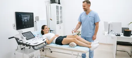
What a knee ultrasound shows
A knee ultrasound shows the structures that lie close to the skin: tendons, the ligaments on the sides of the joint, bursae, muscles and fluid in the joint. The image forms in real time while the doctor moves the probe and bends your knee.
Sound waves bounce off bone and do not pass through it. That makes ultrasound best for the soft tissues around the joint, and for structures deep inside the joint the doctor may recommend an MRI.
The doctor assesses the quadriceps tendon above the kneecap, the patellar tendon below it and both collateral ligaments. They examine the bursae in front of and below the kneecap and on the inner shin (pes anserinus). At the back of the knee they look for a Baker's cyst. They measure the amount of fluid in the joint and the thickness of the joint lining (synovium). The outer edge of the meniscus and cysts next to it are also visible.
Ligaments: what ultrasound can and cannot see
The collateral ligaments lie near the surface, so ultrasound shows them well. The doctor can see whether a ligament has thickened after a sprain, is partly torn or fully ruptured.
The anterior and posterior cruciate ligaments sit in the middle of the joint, between the thigh bone and the shin bone. Ultrasound sees them only partly or indirectly, through effusion and swelling. A reliable assessment of the cruciate ligaments comes from an MRI.
When a knee ultrasound is done
A knee ultrasound is most often requested when the knee swells, hurts when walking, squatting or climbing stairs, or after an injury. Orthopedists often book it as the first imaging test because it is quick and needs no preparation.
The most common reasons for the scan:
- a swollen, tight knee when effusion (fluid on the knee) is suspected
- a lump or fullness behind the knee when a Baker's cyst is suspected
- pain and swelling in front of the kneecap, for example after long kneeling (bursitis)
- pain on the inner side of the knee just below the joint, or below the kneecap when running and jumping
- a twist or a blow to the knee during sport, and follow-up of a healing tendon or ligament
Sports knee injuries
In athletes, ultrasound shows inflammation and partial tears of the patellar tendon (jumper's knee) and the quadriceps tendon. It also shows injury to the inner collateral ligament, which is common in football and skiing, and hamstring strains.
The scan is easy to repeat. At a follow-up the doctor compares the tendon with the earlier images and judges when it is safe to increase the load.
When not to wait for a booked scan
If the knee has suddenly become warm, red and very painful, with a fever, see a doctor the same day. If you notice a swollen calf together with shortness of breath or chest pain, call emergency services at 194 right away.
Preparing for a knee ultrasound
No preparation is needed for a knee ultrasound. Eat, drink and take your medication as usual.
Before you come, it helps to:
- wear shorts or loose trousers that roll up easily above the knee
- bring earlier results: X-ray, MRI or a previous ultrasound
- bring a report from an orthopedist or physiatrist if you have one
- remember when the pain started and which movement triggers it
How the scan works and how long it takes
You lie on your back with the leg straight or a small cushion under the knee. The doctor applies gel and moves the probe over the front and sides of the knee.
For the back of the knee you turn onto your stomach. The doctor then examines the hollow behind the knee and looks for a Baker's cyst.
A knee ultrasound is a dynamic scan. The doctor asks you to bend and straighten the knee or tighten the thigh muscle and watches how the tendon and ligament behave in motion. If needed, the healthy knee is scanned right away for comparison.
The scan usually takes 15 to 30 minutes, and a little longer for both knees. It does not hurt, although a spot that is already sore may be tender under the probe. You can carry on with your day straight after.
What the knee ultrasound report shows
The doctor tells you the main findings during the scan, and you receive the written report shortly after. Reports and images are stored digitally, so a follow-up scan can be compared with the previous one.
A common finding is joint effusion, meaning excess fluid, and the doctor estimates the amount. A Baker's cyst is a fluid-filled sac behind the knee. The doctor measures its size and looks for signs of rupture.
In bursitis the bursa is thickened and filled with fluid. In tendinopathy the tendon is thickened and its structure altered from overload. The scan also shows a partial or complete tear of a tendon or collateral ligament. Thickened synovium may point to joint inflammation.
Based on the findings, the orthopedist suggests treatment: rest, exercises, physiotherapy, medication or further tests. If inflammation markers or uric acid need checking, blood tests are done at the same hospital.
Baker's cyst and a swollen calf
A Baker's cyst usually accompanies another problem in the knee, such as osteoarthritis or meniscus damage. When it ruptures, fluid runs down into the calf and causes swelling and pain similar to deep vein thrombosis.
A knee ultrasound shows the cyst and the fluid around it. If the doctor suspects thrombosis, the veins are checked with a leg vein Doppler.
Knee ultrasound or MRI
Ultrasound and MRI answer different questions, so they often complement each other.
Ultrasound is fast and dynamic. The knee is examined in motion, the painful knee is compared with the healthy one on the spot, and the scan is easy to repeat. Tendons, collateral ligaments, bursae, effusion and a Baker's cyst are clearly visible.
MRI sees the inside of the joint better: the meniscus, cartilage, cruciate ligaments and bone. When a torn meniscus or anterior cruciate ligament is suspected, the orthopedist usually asks for an MRI.
A common order is an orthopedic exam, then a knee ultrasound, and an MRI when the ultrasound does not give the full answer.
Knee ultrasound cost
A knee ultrasound costs from 6,000 RSD. Book your appointment by phone at 062-399-888. When you book, tell us whether one or both knees need scanning. Prices for other diagnostics are in the price list.
- Knee ultrasound
- Soft tissue ultrasound
- Orthopedic consultation
For an appointment call 062-399-888.
Knee ultrasound: frequently asked questions
No. Come as you are, wear clothing that rolls up easily above the knee and bring any earlier results.
Usually 15 to 30 minutes. Scanning both knees takes a little longer, and you can carry on with your day straight after.
No. You feel the gel and light pressure from the probe, and a spot that is already sore may be tender as the doctor passes over it.
Only partly. Ultrasound sees the outer edge of the meniscus and cysts next to it, while the inside of the meniscus and the cartilage are reliably shown by MRI.
For the collateral ligaments, yes, because they lie near the surface. The anterior cruciate ligament is deep in the joint and needs an MRI.
It depends on the complaint. For tendons, bursae, effusion and a Baker's cyst ultrasound is quick and sufficient, while MRI is better for the meniscus, cartilage and cruciate ligaments.
Yes. Ultrasound uses no radiation, so it suits children too, for example for swelling after a fall or pain below the kneecap in children who do sport.
A knee ultrasound costs from 6,000 RSD. Book your appointment by phone at 062-399-888. When you book, tell us whether one or both knees need scanning. Prices for other diagnostics are in the price list.
Related pages
Related exams